 Back to projects

Pulmonary Nodule Diagnosis Based on CT Images
- Date:12/17
- Team: Yi Xu, Xiaohui Lin, Yamin Li, Lanqing Liu, Dayi Li, Wen Gu, Renhui Zhang
- Goal: To achieve intelligent pulmonary nodule diagnosis based on CT, including nodule detection and segmentation, benign-malignant classification, accurate prognosis, image omics.
Brief Introduction
Cancer is one of the greatest threats to people's life and health, among which the incidence rate and mortality rate of lung cancer are the highest. Generally speaking, the 5-year survival rate of early lung cancer is more than 50% higher than that of advanced lung cancer. Therefore, the early diagnosis of lung cancer is very important for saving the lives of patients. Because early lung cancer has fewer clinical symptoms and is difficult to screen, lung nodule detection based on CT imaging analysis has become an important means for early diagnosis of lung cancer.
To solve this problem, we develop a 3D pulmonary nodule detection algorithm based on CT image analysis, which mainly consists of two processes: generation of candidate lung nodule areas and false positive reduction. What’s more, we work closely with Shanghai Chest Hospital, and a chest CT database with more than 3,000 images is expected to be established. So far, the overall sensitivity and average sensitivity of our algorithm are 98.7% and 84.2% on LUNA16 dataset.
Our follow-up works will focus on accurate prognosis of pulmonary nodules and image omics. The former task mainly contains differentiation of benign and malignant pulmonary nodules, intelligent follow-up including the prediction of pulmonary nodule doubling time, etc. For the latter task, we will develop nodule image feature analysis and find its relationship with pathological characteristics and immunohistochemistry.
Nodule detection algorithm:

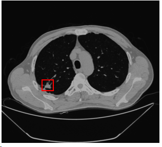
Part of detection results:

Paper
Coming soon!
Dataset
Coming soon!
